
Julien PRUDENT
Mitochondrial Dynamics lab
Published on 06.10.2026
Presentation

Understanding mitochondrial dynamics, organelle communication and mitochondrial DNA signalling
Mitochondria are highly dynamic organelles that continuously remodel their membranes and communicate with other cellular compartments. Beyond their central role in energy production, mitochondria act as major signalling hubs controlling cellular metabolism, calcium homeostasis, cell death, inflammation and immunity. Defects in mitochondrial function and morphology are associated with a broad range of human diseases, from primary mitochondrial disorders to neurodegenerative diseases and cancer.Our laboratory investigates the molecular mechanisms that shape mitochondrial architecture and morphology and determine how mitochondria communicate with the rest of the cell. We are particularly interested in the interplay between mitochondrial membrane remodelling, mitochondria–organelle contact sites and mitochondrial DNA (mtDNA) dynamics, and in understanding how these processes influence cellular homeostasis and disease.
Our research combines advanced live-cell and super-resolution microscopy, electron microscopy, and quantitative cell biology to investigate mitochondrial behaviour, as well as employing biochemical, molecular and proteomic approaches to identify the molecular machinery controlling these processes.
Area 1: Mitochondrial membrane dynamics
Mitochondria constantly undergo fusion and division, allowing their network to adapt to changes in cellular metabolism and stress. These membrane-remodelling events are essential for mitochondrial quality control, the distribution of mitochondrial components and the regulation of cell fate. Our laboratory aims to uncover the molecular mechanisms controlling mitochondrial membrane dynamics, and how inter-organellar interactions control mitochondrial division and fusion.
Our work has identified novel regulators of mitochondrial morphology and has revealed an unexpected contribution of Golgi-derived vesicles to mitochondrial membrane remodelling. We showed that Golgi-derived vesicles carrying specific phosphoinositides are recruited to mitochondrial constriction sites and participate in mitochondrial division. These observations also uncovered complex multi-organelle interfaces involving mitochondria, the endoplasmic reticulum, lysosomes and Golgi-derived membranes.
We are now investigating additional regulators of membrane remodelling, as well as how these organelle interactions coordinate mitochondrial fission and fusion, and how disruption of these pathways affects mitochondrial function, metabolism and cell survival.
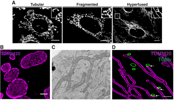
Figure 1. Mitochondrial membrane dynamics. Representative mitochondrial morphologies and super-resolution and electron microscopy of mitochondrial architecture (A-C), and membrane contact sites between mitochondria and Golgi-derived vesicles are visualised by super-resolution microscopy.
Axe 2 : Mitochondria–organelle contact sites
Mitochondria do not function in isolation, but establish close membrane contacts with multiple other cellular compartments, including the endoplasmic reticulum, lysosomes, endosomes, peroxisomes, lipid droplets, and Golgi-derived vesicles. These mitochondria–organelle contact sites create specialised signalling platforms through which organelles exchange lipids, metabolites, calcium and signals. They therefore provide an important mechanism for coordinating the activity of different cellular compartments.Our goal is to identify the proteins and molecular pathways that establish and regulate these contacts and to determine how different organelles cooperate at multi-organelle interfaces. Using high-resolution and live-cell imaging together with proteomic and biochemical approaches, we investigate how membrane contact sites regulate mitochondrial architecture, metabolism, signalling and cellular stress responses, and how their dysfunction contributes to human diseases.
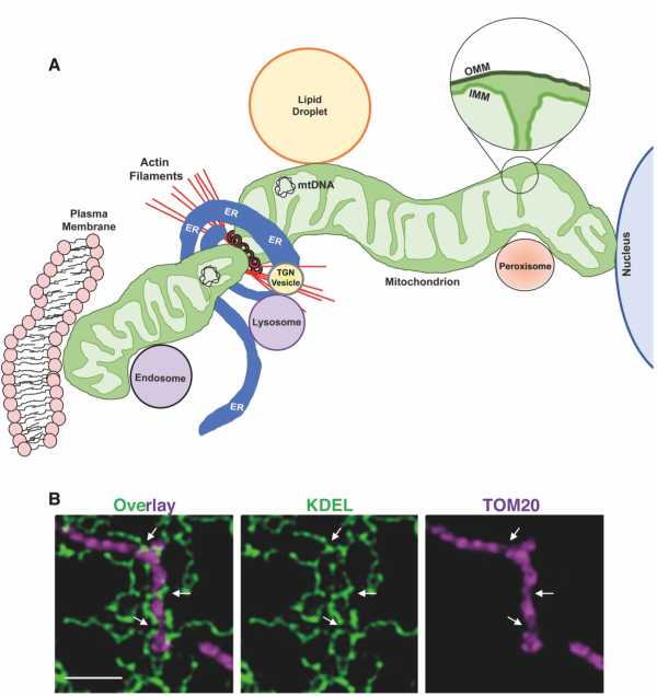
Figure 2. Mitochondria–organelle contact sites. Mitochondria establish membrane contact sites with multiple cellular organelles. Super-resolution microscopy illustrates contacts between mitochondria and the endoplasmic reticulum.
Axe 3 : Mitochondrial membrane remodelling and mitochondrial DNA
The mitochondrial genome is packaged into nucleoids distributed throughout the mitochondrial network, and its maintenance is closely linked to mitochondrial membrane dynamics. However, how mitochondrial architecture controls mtDNA distribution, quality control and fate remains incompletely understood.Our work has revealed that mitochondrial membrane remodelling plays an important role in mtDNA life cycle. We identified an MTFP1-dependent pathway that isolates damaged inner mitochondrial membrane compartments for autophagic degradation and contributes to basal and stress-induced mtDNA turnover. Conversely, we showed that mitochondrial stress induced by fumarate accumulation promotes the formation and release of mtDNA-containing mitochondrial-derived vesicles, activating innate immune signalling.
Our laboratory aims to understand how mitochondrial dynamics and organelle interactions determine whether mtDNA is maintained, degraded or released from mitochondria, and how disruption of these processes contribute to inflammatory signalling and diseases.
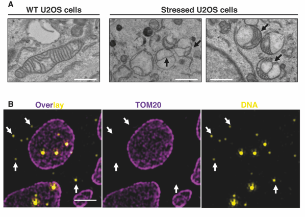
Figure 3. Mitochondrial membrane remodelling and mtDNA dynamics. Electron and super-resolution microscopy show mitochondrial membrane remodelling and mitochondrial DNA dynamics following mitochondrial stress.