Sigolène Meilhac
Heart Morphogenesis
Published on 23.04.2026
Presentation

The Imagine-Pasteur group of Heart Morphogenesis studies how cells are patterned and how coordinated cell behaviour generates 3D tissue deformations to shape the organ. We address these questions using cutting-edge technologies in genetics, omics, 3D imaging, quantitative analyses of complex data and computer simulations. Our study meets novel challenges requiring interdisciplinary efforts in collaboration with computational scientists and clinicians, to quantify biological processes or examine the clinical impact of fundamental knowledge. In the last years, we have developed several tools to image, simulate and quantify heart development and malformation in 3D. Our work in the mouse provides conceptual advances in developmental biology, as well as novel perspectives in pediatric cardiology.

- Left-right asymmetry of the heart
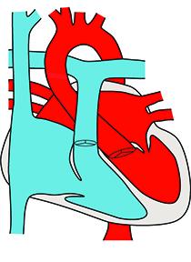
The heart is an asymmetric organ, in which the left and right sides are fully separated to drive the double blood circulation in mammals. The molecular cascade breaking bilateral symmetry in the early embryo has been well characterised, with the formation of a left-right organiser, the node. However, how left-right asymmetric gene expression is later sensed by organ specific precursor cells to generate asymmetric morphogenesis is poorly understood. Our aim is to dissect how patterning of heart precursors drives asymmetric heart morphogenesis, and how anomalies in this process are associated with congenital heart defects.
In the early embryo, looping of the heart tube, is the first morphological sign of left-right asymmetry. It corresponds to the transformation of the initial straight heart tube into a helix. Heart looping is essential to align the cardiac chambers and great vessels and thus establish the plumbing of the blood flow. Previously, heart looping had been described as a binary process (rightward or leftward direction), corresponding to the readout of the symmetry-breaking event, but the fine 3D shape of the heart loop, as a readout of asymmetric morphogenesis had not been addressed. We thus propose a paradigm shift in the analysis of heart looping, from a binary process to spatio-temporal dynamics. This will be essential to uncover the pathophysiology of laterality defects such as heterotaxy, which are associated with a broad phenotypic variability.
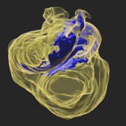
Using High Resolution Episcopic Microscopy (HREM), we have now reconstructed mouse heart looping dynamics in 3 dimensions and developed tools to quantify the shape of the heart tube, not just its direction. The staging criteria that we have established provide a higher temporal resolution of early mouse heart development. We have proposed a novel mechanism of heart looping and developed a computer model, to predict the shape of the looped heart tube from initial mechanical constraints and sequential left-right asymmetries (Le Garrec et al., 2017).
Recently, we have analysed how left-sided Nodal signalling, which is impaired in the heterotaxy syndrome, shapes the embryonic heart tube. We have shown that Nodal is not required to initiate heart looping, but rather to amplify and orient left-right asymmetries at the tube poles. We have dissected the spatio-temporal window of Nodal signalling in heart precursors, as well as its downstream effectors (Desgrange et al., 2020).
Our work on mouse heart morphogenesis is relevant to congenital heart defects in humans, such as misalignment of cardiac chambers. Congenital heart diseases represent a major concern for public health, affecting 1% of newborns and a leading cause of death in utero and during the first year of life. However, the genetic bases of these defects and the underlying pathophysiological processes remain poorly understood.
- Mechanism of heart growth
Growth of the heart is required to adapt to the increasing hemodynamic load of the developing organism. Physiologically, heart growth is mainly driven by the proliferation of myocardial cells in utero and by cell size increase after birth. However, how myocardial cells cease to proliferate after birth remains poorly understood. In addition to growing in size, the myocardium matures in its architecture, with the alignment of cardiomyocytes into fibres. This orientation is an important aspect of the efficient heart contraction. A current question is to understand how the architecture of the myocardium is established.
We have previously characterised, by clonal analysis, the lineages and behaviour of myocardial cells during heart morphogenesis. We have developed interdisciplinary tools for the quantification of tissue anisotropy in 3D and revealed that myocardial cells coordinate locally their orientation of division during cardiac chamber expansion. We have studied the atypical cadherin Fat4, which was initially discovered in the fly as a major regulator of organ size. However, how the Fat pathway is connected to the Hippo pathway in mammals had remained poorly understood. We have shown that Fat4 is required to restrict heart growth at birth, by modulating the nuclear translocation of an adaptor protein, Amotl1, which is a partner of an effector of the Hippo pathway, Yap1.
Our work on mouse heart morphogenesis is relevant to heart repair in humans. Heart failure is the leading cause of death in the industrialized world, owing to the limited regenerative capacity of the mammalian heart. In addition to controlling the number of cardiomyocytes, it will be important to promote their oriented arrangement for producing a mature myocardium with efficient contraction.
Team


Resources & publications
-
 2023Journal (source)Dev Cell
Identification of Greb1l as a genetic determinant of crisscross heart in mice...
-
2021Journal (source)Annu Rev Gen Hum Genet
Heart Development and Congenital Structural Heart Defects.
-
2020Journal (source)Dev Cell
Transient Nodal Signaling in Left Precursors Coordinates Opposed Asymmetries ...
-
Journal (source)Philos Trans R Soc Lond B Biol Sci
Mesoderm patterning by a dynamic gradient of retinoic acid signalling.
-
2020Journal (source)Cell Rep
Intraflagellar Transport Complex B Proteins Regulate the Hippo Effector Yap1 ...
-
2019Journal (source)Dis Model Mech
Standardised imaging pipeline for phenotyping mouse laterality defects and as...
-
2018Journal (source)Development
Left-right asymmetry in heart development and disease: forming the right loop.
-
2018Journal (source)Nat Rev Cardiol
The deployment of cell lineages that form the mammalian heart.
-
2017Journal (source)Elife
A predictive model of asymmetric morphogenesis from 3D reconstructions of mou...
-
2017Journal (source)Am. J. Hum. Genet.
Mutations in GREB1L Cause Bilateral Kidney Agenesis in Humans and Mice.
-
2017Journal (source)Nat Commun
Amotl1 mediates sequestration of the Hippo effector Yap1 downstream of Fat4 t...
-
Journal (source)Cold Spring Harb Perspect Med
Cardiac cell lineages that form the heart.
-
2015Journal (source)Cilia
The more we know, the more we have to discover: an exciting future for unders...
-
Journal (source)Wiley Interdiscip Rev Syst Biol Med
Integrating multi-scale knowledge on cardiac development into a computational...
-
2014Journal (source)Med Sci (Paris)
[Cilia and heart morphogenesis].
-
2013Journal (source)Bioinformatics
Extracting 3D cell parameters from dense tissue environments: application to ...
-
2013Journal (source)Dev. Dyn.
Resolving cell lineage contributions to the ventricular conduction system wit...
-
2013Journal (source)Development
Quantitative analysis of polarity in 3D reveals local cell coordination in th...
-
2012Journal (source)Circ. Res.
Asymmetric fate of the posterior part of the second heart field results in un...
-
Journal (source)Results Probl Cell Differ
Cell lineages, growth and repair of the mouse heart.
-
2011Journal (source)Dev. Cell
Tracing cells for tracking cell lineage and clonal behavior.
-
2010Journal (source)Development
Clonal analysis reveals common lineage relationships between head muscles and...
-
2010Journal (source)Circ. Res.
Biphasic development of the mammalian ventricular conduction system.
-
2009Journal (source)Dev. Biol.
Active cell movements coupled to positional induction are involved in lineage...
-
2009Journal (source)Circ. Res.
Landmarks and lineages in the developing heart.
-
2008Journal (source)Dev. Biol.
Myocardium at the base of the aorta and pulmonary trunk is prefigured in the ...
-
2007Journal (source)BMC Dev. Biol.
Regionalization of the mouse visceral endoderm as the blastocyst transforms i...
-
2007Journal (source)Dev. Biol.
The anterior visceral endoderm of the mouse embryo is established from both p...
-
2006Journal (source)Dev. Biol.
Left and right ventricular contributions to the formation of the interventric...
-
2006Journal (source)Development
Smooth muscle of the dorsal aorta shares a common clonal origin with skeletal...
-
2005Journal (source)Nat. Rev. Genet.
Building the mammalian heart from two sources of myocardial cells.
-
2005Journal (source)Circ. Res.
Right ventricular myocardium derives from the anterior heart field.
-
2004Journal (source)Dev. Cell
The clonal origin of myocardial cells in different regions of the embryonic m...
-
2004Journal (source)J. Cell Biol.
Oriented clonal cell growth in the developing mouse myocardium underlies card...
-
2003Journal (source)Development
A retrospective clonal analysis of the myocardium reveals two phases of clona...

Key numbers
34 publications